Our Platform · AI Toxicity
Our AI turns organoid images into consistent toxicity calls
From image upload to toxicity call
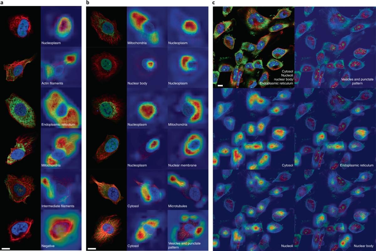
The software analyzes live/dead fluorescence and time-lapse brightfield images, learns how they change over time, and predicts drug response. The same criteria apply to every image, and hundreds of images are processed in one run.
Well detection and quality check
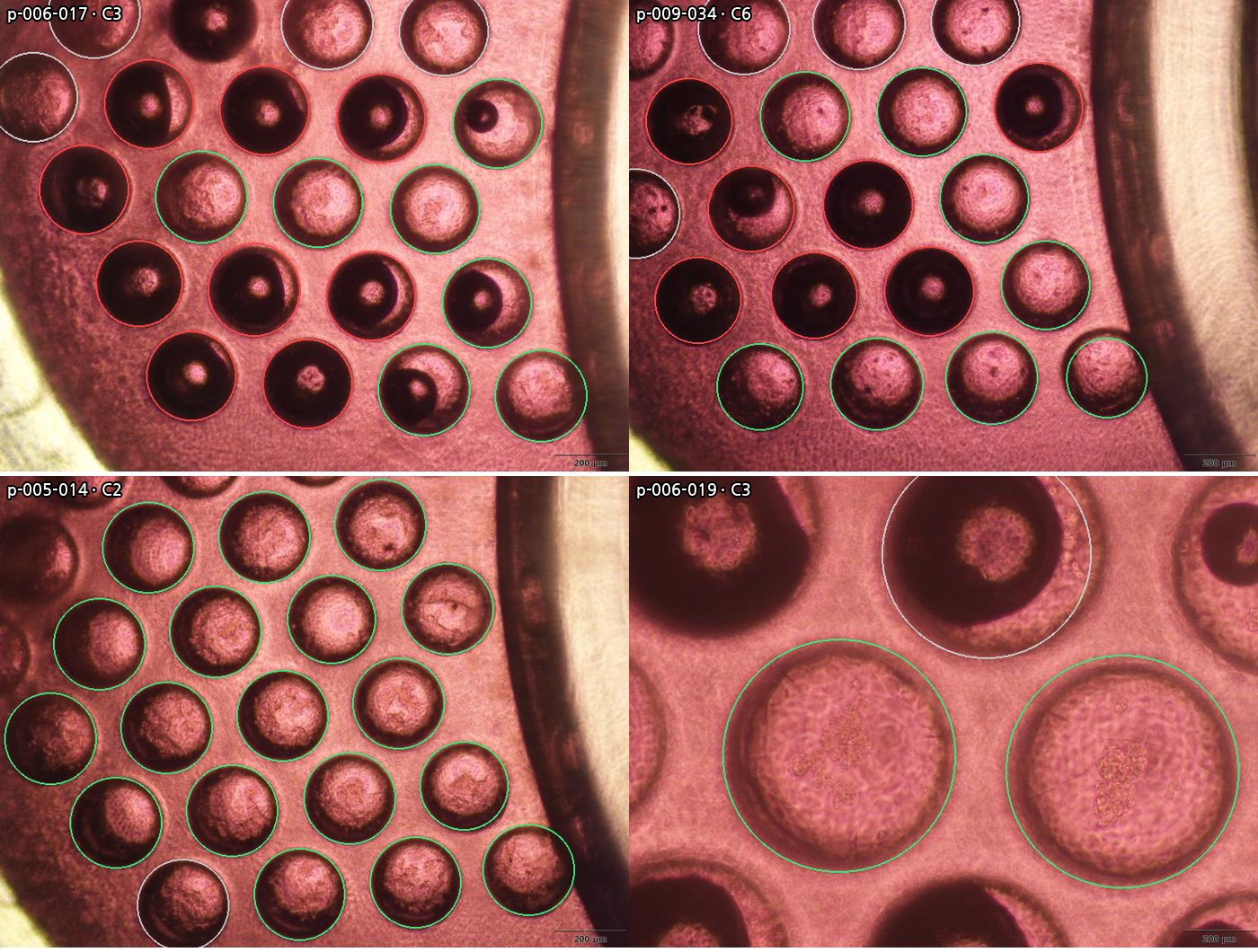
Finds Port Pocket microwells at any magnification and excludes wells blocked by bubbles or unfilled media.
Segmentation
Segments organoids in brightfield and fluorescence images and measures area, shape and live/dead signal per well.
Early toxicity prediction
Uses early time points to predict the endpoint toxicity grade (T0, T1, T2) and viability.
Labeling, retraining and reports
Corrections become training data. Results export as PDF and Excel with a full audit trail.
Validated on AiderBio images
We tested well detection and readout on 29 Port Pocket images containing 229 microwells. Readout accuracy was measured on culture conditions that were not used to set the model.
Reference labels by one AiderBio reader; confirmation by two or more readers is planned.
Model tracks
Track 1 · Anomaly detection
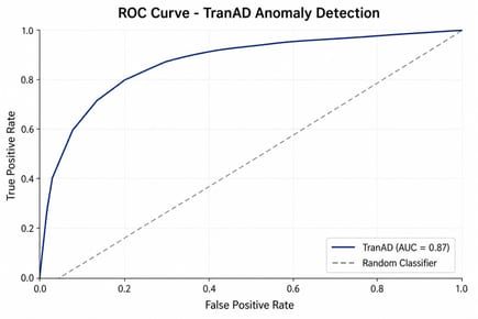
A transformer-based time-series model reads day 1–7 brightfield images (shape, size, density) and flags when 3D growth becomes abnormal.
Track 2 · Viability prediction
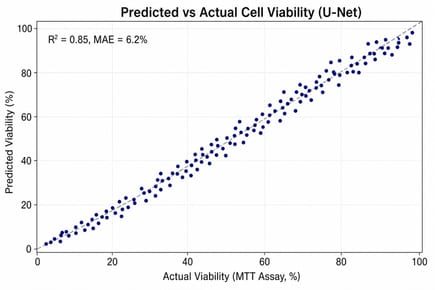
A U-Net model learns from Calcein-AM time-lapse images paired with MTT viability and predicts viability from images alone.